About this deal
Huston, J. P. (1982). “Searching for the neural mechanism of reinforcement (of ‘stamping in’),” in The Neural Basis of Feeding and Reward, eds B. G. Hoebel and D. Novin (Florida, Fl: Academic Press), 75–83. Huston, J. P., and Borbély, A. A. (1973). Operant conditioning in forebrain ablated rats by use of rewarding hypothalamic stimulation. Brain Res. 50, 467–472. doi: 10.1016/0006-8993(73)90753-1 Candy, B., Jones, L., Williams, R., Tookman, A., and King, M. (2008). Psychostimulants for depression. Cochrane Datab. Systemat. Rev. 2008:CD006722. doi: 10.1002/14651858.CD006722.PUB2/INFORMATION/EN Janssen, N. P., Hendriks, G. J., Baranelli, C. T., Lucassen, P., Voshaar, R. O., Spijker, J., et al. (2021). How Does Behavioural Activation Work? A Systematic Review of the Evidence on Potential Mediators. Psychother. Psychosomat. 90, 85–93. doi: 10.1159/000509820 Edmonds, D. E., and Gallistel, C. R. (1977). Reward versus performance in self-stimulation: electrode-specific effects of alpha-methyl-p-tyrosine on reward in the rat. J. Comp. Physiol. Psychol. 91, 962–974. doi: 10.1037/h0077391
The birth, death and resurrection of avoidance: a - Nature
Gallistel, C. R. (1978). Self-stimulation in the rat: quantitative characteristics of the reward pathway. J. Comp. Physiol. Psychol. 92, 977–998. doi: 10.1037/h0077513 In the following subsections we summarize evidence that gave rise to the series-circuit hypothesis as well as evidence that challenges this longstanding account of brain-reward circuitry. We then discuss the implications of the convergence model for interpretation of the effect of MFB stimulation on relief of treatment-resistant depression. Intracranial Self-Stimulation of the Medial Forebrain Bundle: Phenomenology One such action circuitry is the loop between prefrontal cortex and basal ganglia which is involved in gaze control 8, 9. Prefrontal cortex is known to be sensitive to object novelty 10, 11 even from childhood 12. Prefrontal cortex is also implicated in processing rewarding and aversive stimuli 13, 14, 15. Likewise selective coding of rewarding and/or punishing stimuli is observed in basal ganglia 3, 16, 17. In addition, novel objects are also known to invoke enhanced responses in areas within basal ganglia such as in caudate 18. We have previously shown that value memories shape the visual responses of objects similarly and with a remarkable granularity in vlPFC and the basal ganglia output, SNr. 19. However, it is not known whether the visual responses to objects in the corticobasal circuitry are also affected by other past experiential dimensions such as perceptual exposure (familiarity vs novelty) or aversive associations and if so whether the same neurons encode such disparate object memories across different domains.Coenen, V., Hurwitz, T., Panksepp, J., Mädler, B., and Honey, C. (2009b). Medial forebrain bundle stimulation elicits psychotropic side effects in Subthalamic Nucleus Deep Brain Stimulation for PD – new insights through Diffusion Tensor Imaging. Akt Neurol. 36, s–0029–1238842. doi: 10.1055/s-0029-1238842 The animal study that generated the publicly available dataset was reviewed and approved by the Animal Research Ethics Committee, Concordia University. Author Contributions Kim, K. M., Baratta, M. V., Yang, A., Lee, D., Boyden, E. S., and Fiorillo, C. D. (2012). Optogenetic mimicry of the transient activation of dopamine neurons by natural reward is sufficient for operant reinforcement. PLoS One 7:e33612. doi: 10.1371/journal.pone.0033612 Amsterdam, J. D., Settle, R. G., Doty, R. L., Abelman, E., and Winokur, A. (1987). Taste and smell perception in depression. Biol. Psychiat. 22, 1481–1485. doi: 10.1016/0006-3223(87)90108-9
Jedec Waffle Trays | Action Circuits | Products
vlPFC was found to be sensitive to all three domains of object memory including reward, aversiveness, mere exposure (novelty/familiarity). Importantly, these domains were not encoded by separate populations rather they were encoded by the same neurons and in a correlated fashion. The degree and sign of responding were consistent with the ecological salience of the objects measured during free viewing (Supplementary Figs. 4, 6, 8). While in this study, we only considered long-term familiarity and absolute novelty, previous evidence showed the same vlPFC neurons to have significant suppression to recently viewed objects (relative novelty aka recency) 5, 19. Recency is also shown to reduce objects’ ecological salience 5. Together these results suggest vlPFC to encode a common currency signal related the attention worthiness of objects. This is consistent with the role of vlPFC in controlling gaze via anatomical projections to frontal eye field (FEF) 29 and superior colliculus (SC) 9. Wiegert, J. S., Mahn, M., Prigge, M., Printz, Y., and Yizhar, O. (2017). Silencing Neurons: Tools, Applications, and Experimental Constraints. Neuron 95, 504–529. doi: 10.1016/j.neuron.2017.06.050 Guyenet, P. G., and Aghajanian, G. K. (1978). Antidromic identification of dopaminergic and other output neurons of the rat substantia nigra. Brain Res. 150, 69–84. doi: 10.1016/0006-8993(78)90654-6 Olds, J. (1973). “The discovery of reward systems in the brain,” in Brain stimulation and motivation: research and commentary, ed. E. S. Valenstein (Glenview, IL: Scott, Foresman), 81–99. Maeda, H., and Mogenson, G. J. (1980). An electrophysiological study of inputs to neurons of the ventral tegmental area from the nucleus accumbens and medial preoptic-anterior hypothalamic areas. Brain Res. 197, 365–377. doi: 10.1016/0006-8993(80)91122-1Deutsch, J. A., Adams, D. W., and Metzner, R. J. (1964). Choice of intracranial stimulation as a function of delay between stimulations and strength of competing drive. J. Comparat. Physiol. Psychol. 57, 241–243. doi: 10.1037/h0047915 Pound, P., and Ritskes-Hoitinga, M. (2018). Is it possible to overcome issues of external validity in preclinical animal research? Why most animal models are bound to fail. J. Transl. Med. 16:304. doi: 10.1186/S12967-018-1678-1 Molendijk, M. L., and de Kloet, E. R. (2015). Immobility in the forced swim test is adaptive and does not reflect depression. Psychoneuroendocrinology 62, 389–391. doi: 10.1016/J.PSYNEUEN.2015.08.028 Jbabdi, S., Sotiropoulos, S. N., Haber, S. N., Van Essen, D. C., and Behrens, T. E. (2015). Measuring macroscopic brain connections in vivo. Nat. Neurosci. 18, 1546–1555. doi: 10.1038/nn.4134 Edmonds, D. E., and Gallistel, C. R. (1974). Parametric analysis of brain stimulation reward in the rat: III. Effect of performance variables on the reward summation function. J. Comp. Physiol. Psychol. 87, 876–883. doi: 10.1037/h0037217
Baking and Vacuum packing - Action Circuits Baking and Vacuum packing - Action Circuits
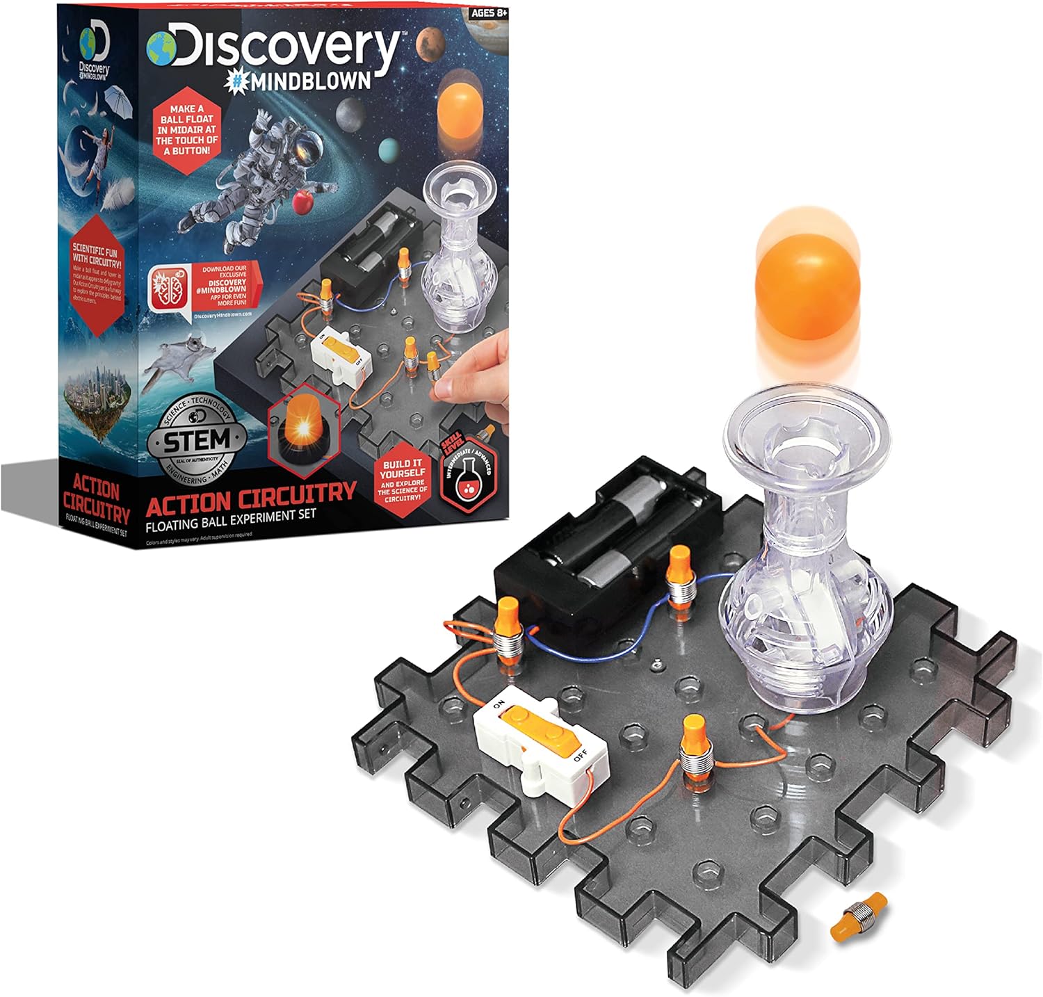
Action Circuits offer a comprehensive electronic component testing service for Semiconductor devices. The failure of psychomotor stimulants to serve as an effective monotherapy for depression invites reconsideration of a series-circuit model of the antidepressant effect of MFB stimulation. An alternative, analogous to the convergence model of intracranial MFB self-stimulation, would include multiple, convergent pathways. On that view, non-dopaminergic MFB components may contribute to the therapeutic effect in parallel to, in synergy with, or even instead of, a dopaminergic component. To assess those possibilities, we must look in more detail at the neuroanatomical complexity of the region where MFB stimulation is effective in relieving treatment-resistant depression and at the methods that have been used to link that effect to particular fiber bundles. Which Neurons Are Activated Directly by Therapeutically Effective Stimulation of the Medial Forebrain Bundle, and Which Are Responsible for the Antidepressant Effect? Once the trajectories of the axons of interest have been traced, optogenetic methods ( Yizhar et al., 2011) can render the neurons that give rise to particular MFB components optically excitable, thereby making it possible to determine whether driving these cells produces rewarding and/or motivating effects. Identification of the terminal fields of the MFB-projecting neurons, coupled with optogenetic silencing methods ( Yizhar et al., 2011; Wiegert et al., 2017) provide complementary means for assessing the necessity of these neurons for the rewarding effect of MFB stimulation. By recording the activity of these neurons in response to rewarding MFB stimulation, it can be determined whether the properties of their axons correspond to the psychophysically derived portrait of the fibers subserving MFB self-stimulation (e.g., Rompré and Shizgal, 1986; Shizgal et al., 1989; Murray and Shizgal, 1996b; Cossette et al., 2016). Implications for Research on the Antidepressant Effect of Deep-Brain Stimulation Stimulation Parameters POWERED BY BATTERIES: Each experiment requires 2 AA batteries (12 total, not included) to power the modules. You must connect batteries to run the experiments.Kopnisky, K. L., and Hyman, S. E. (2002). “MOLECULAR AND CELLULAR BIOLOGY OF ADDICTION,” in Neuropsychopharmacology: The Fifth Generation of Progress, eds K. L. Davis, D. Charney, J. T. Coyle, and C. Nemeroff (Philadelphia, PA: Lippincott, Williams, & Wilkins), 1367–1379. doi: 10.1016/j.semcdb.2009.03.002
 Great Deal